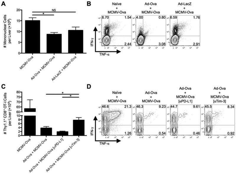
Fig 5.

Tim-3 blockade improves antigen-specific hepatic secondary immune responses to viral infection. C57BL/6 mice were IV infected with 2.5 × 10⁷ IU of Ad-Ova, 2.5 × 10⁷ IU of Ad-LacZ, or left uninfected. At day 7, all three experimental groups were IV infected with 1 × 10⁴ IU of MCMV-Ova. (A) The number of live MNCs isolated from livers of day 14 infected animals was enumerated. Trypan blue exclusion was used to assess the number of viable cells. (B) Endogenous CD8+ T cell TNF-α and IFN-γ were quantified at day 14 after a 5-hour restimulation with 2 μg/mL of SIINFEKL peptide (n = 3 per group). (C and D) In a parallel experiment, 5 × 10⁵ naïve Thy1.1+CD8+ OT-I T cells were transferred at day 7. C57BL/6 mice were left untreated or were administered 300 μg of anti-PD-L1 Ab or anti-Tim-3 Ab IP at days 5 and 6 before a day 7 MCMV-Ova infection. The number of Thy1.1+CD8+ OT-I T cells and their TNF-α and IFN-γ production was assessed in livers of infected animals at day 14. Cytokine detection was achieved after a 5-hour restimulation with 2 μg/mL of SIINFEKL peptide (one-way ANOVA/Tukey's posttest; n = 3 per group). Numbers in the scatter plots represent percentages (mean ± standard error of the mean). *P < 0.05.
