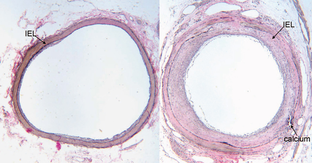
Figure 5.
Representative examples of atherosclerotic lesion from cross sections of the left anterior descending artery of a nondepressed (left) and depressed (right) monkey. Atherosclerotic lesion was quantified as the area of the intima (IA), i.e., the area internal to the internal elastic lamina (IEL). Sections were stained with Verhoeff van Gieson stain. Lesion area of the nondepressed monkey was minimal (IA = 0.08 mm2), consisting of eccentric fatty streak and small fibro-fatty plaque. The lesion of the depressed monkey was much more extensive (IA = 0.52 mm2), consisting of a moderately advanced concentric fibro-fatty to fibro-muscular plaque with a small focus of calcium. Multifocal destruction of the IEL and media and lipid infiltration into the media were apparent.

