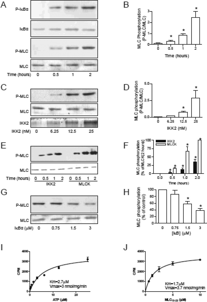
Figure 1. IKK2 phosphorylates MLC in vitro.
A and B, MLC (0.75 µM) or IκBα (0.75 µM) was incubated with IKK2 (25 nM) in a total of 50 µl reaction buffer at 37°C for the indicated time. MLC or IκBα was visualized by ponseau s solution, and the phosphorylated MLC or IκBα was analyzed by western blotting. n=5. *P<0.05 vs. 0; One way ANOVA. C and D, the indicated amount of IKK2 were incubated with MLC (0.75 µM) at 37°C for one hour. n=3. *P<0.05 vs. 0; One way ANOVA. E and F, MLC (0.75 µM) were phosphorylated by IKK2 (25 nM) or MLCK (25 nM) for the indicated time. n=3. *P<0.05 vs. 0; # P<0.05 v.s. MLCK; Two way ANOVA. G and H, MLC (0.75 µM) and IKK2 (25 nM) were incubated at 37°C for one hour in the presence of IKK2 peptide substrate (DSGLDSM). n=3. *P<0.05 vs. 0; One way ANOVA. I and J, kinetic analysis was performed with varying concentrations of ATP (I) or substrate MLC10–23 peptide (J).

