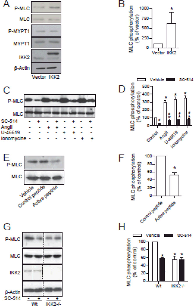
Figure 2. IKK2 phosphorylates MLC in HVSMCs.
A and B, HVSMCs were transfected with flag-tagged IKK2 or vector. MLC phosphorylation and flag-tagged IKK2 expression were analyzed by western blotting. n=3. *P<0.05 vs. vector; student t-test. C and D, after 15 minutes of incubation with SC-514 (30 µM) or vehicle, HVSMCs were treated with the indicated agonist for 3 minutes (angiotensin II, 0.1 µM) or 20 minutes (U-46619, 1 µM and ionomycin, 1 µM), MLC phosphorylation was analyzed by western blotting. n=3. *P<0.05 vs. control; # P<0.05 vs. vehicle; one way ANOVA. E and F, HVSMCs were treated with inhibitory peptide (A 14-amino acid peptide corresponding to the active IκB phosphorylation recognition sequence fused to the hydrophobic region of the fibroblast growth factor signal peptide to aid in cellular delivery) or control peptide [A 14-amino acid peptide corresponding to the mutated recognition sequence of IκB (Ser32→Ala and Ser36→Ala) fused to the hydrophobic region of the fibroblast growth factor signal peptide to aid in cellular delivery] for 30 minutes, MLC phosphorylation was then analyzed by western blotting. n=3. *P<0.05 vs. control; student t-test. G and H, wild-type and IKK2−/− MEFs were incubated with SC-514 (30 µM) for 15 minutes, MLC phosphorylation and IKK2 expression in wild-type and IKK2−/− MEFs was analyzed by western blotting. n=3. *P<0.05 vs. control; student's t-test.

