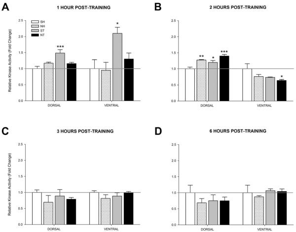
Figure 6.
Activity levels of PKA at 1, 2, 3, or 6 hours post-training or injection (n=4 for each group). Mice either received acute injections of saline (SH) or nicotine (0.09 mg/kg; NH) then were left in their home cages or acute injections of saline (ST) or nicotine (0.09 mg/kg; NT) then trained in contextual conditioning. Bilateral dorsal and ventral hippocampi were then removed (A) 1 hour post-training, (B) 2 hours post-training, (C) 3 hours post-training, or (D) 6 hours post-training and then subjected to the PKA activity assay. Data are presented as fold change in kinase activity relative to the respective SH group. * p < 0.05, ** p < 0.01, *** p < 0.001 compared to the SH group only. Data are presented as mean ± SEM.

