Figure 1.

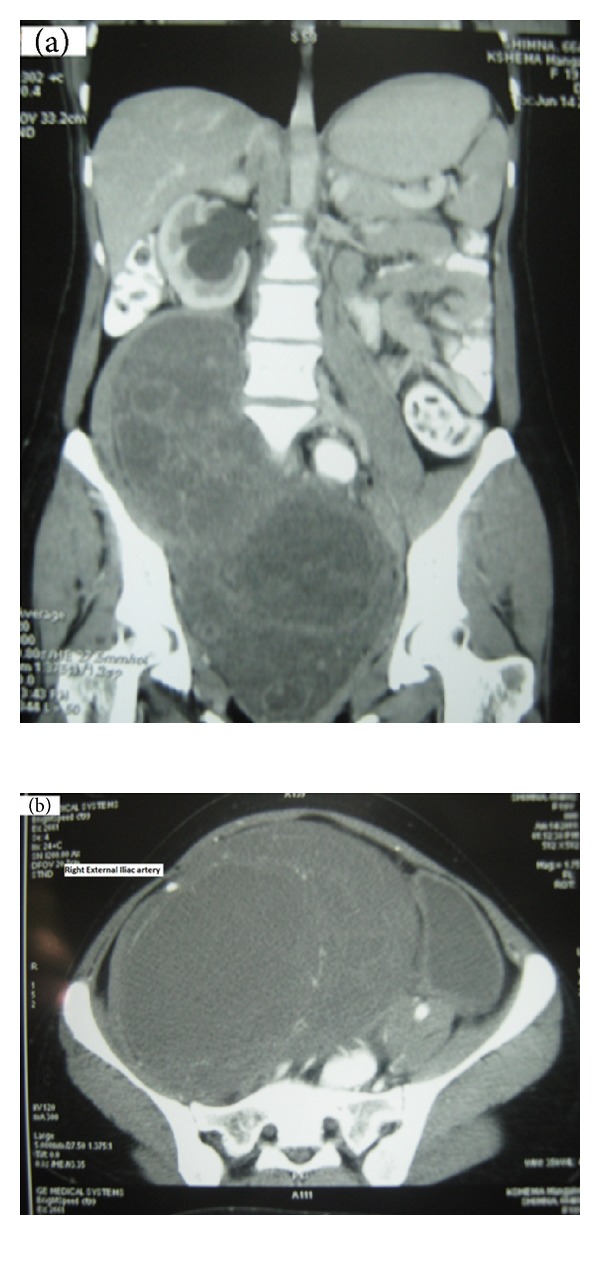
(a) C.T. scan showing large lobulated mass lesion in the pelvic retroperitoneum. (b) Right external iliac artery stretched over the mass.

(a) C.T. scan showing large lobulated mass lesion in the pelvic retroperitoneum. (b) Right external iliac artery stretched over the mass.