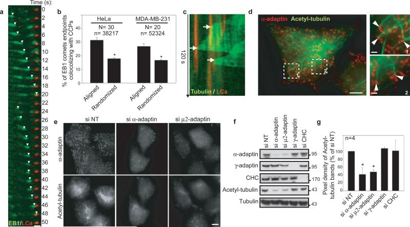
Figure 1. Microtubules pause at CCPs and are acetylated in an AP-2-dependent manner.
a, b, GFP-EB1 comets stopping at CCPs (a, TIRF-M, HeLa cells) and quantification (b, see Method section; number of cells (N) and EB1 comets (n)). c, GFP-tubulin-positive microtubule contacting CCP. d, e, Control (d) or siRNAs-treated (e) HeLa cells stained for α-adaptin and K40 acetyl-tubulin. f, g, Proteins expression in HeLa cells treated with indicated siRNAs (molecular weights in kDa). Quantification in percentage ± s.e.m of siNT, * P<0.001. Scale bars, 10 μm or 2 μm in insets.

