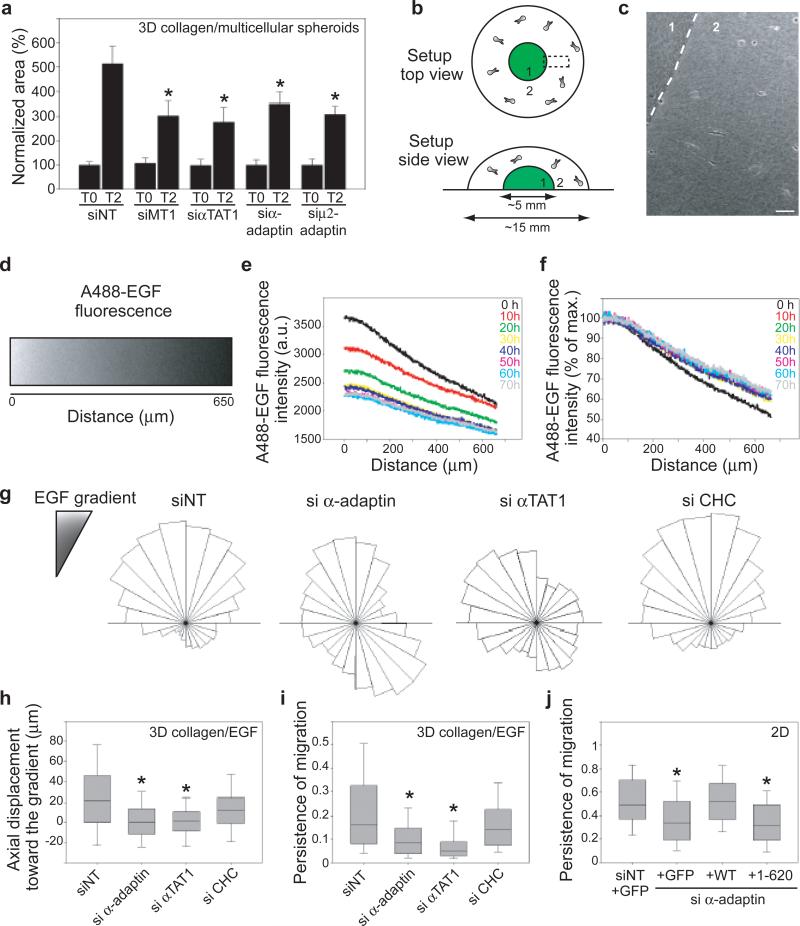
Figure 4. αTAT1 interaction with AP-2 is required for directed cell migration.
a, Area of 3D invasion after 2 days (T2) by spheroids of siRNAs-treated MDA-MB-231 cells. b, c Schematic representation (b) or phase-contrast image (c) of the 3D collagen I EGF-chemotaxis setup. Scale bar, 50 μm. d, A488-EGF gradient at time 0 in a region corresponding to boxed area in (b). e-f, Evolution over time of the intensity (e) and slope (f) of the A488-EGF gradient. g, Angular distribution relative to gradient orientation of siRNAs-treated MDA-MB-231 cells. h-i, Axial displacement towards the gradient (h) and persistence of migration (i) of siRNAs-treated MDA-MB-231 cells. j, Persistence of migration on glass coverslip of MDA-MB-231 cells depleted for α-adaptin and expressing the indicated constructs. Error bars indicate mean ± s.e.m (*P<0.001 in a and P<0.05 in h-j).

