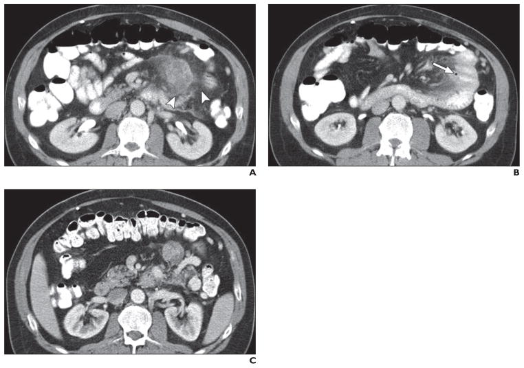
Fig. 10. 59-year-old woman with acute abdominal pain and pancreatic neuroendocrine tumor and mesenteric nodal metastatic lesions treated with sunitinib for 7 months.
A, Axial contrast-enhanced CT image obtained at acute presentation shows haziness (arrowheads) around mesenteric mass and adjacent jejunal loop.
B, Axial contrast-enhanced CT image obtained at same examination as A at more inferior level shows small sealed perforation along mesenteric border of proximal jejunal loop (arrow). These findings were regarded as complication of sunitinib treatment, which include small-bowel ischemia and perforation. Patient was treated conservatively with antibiotics, and sunitinib treatment was stopped.
C, Axial contrast-enhanced CT image obtained 1 month after A and B shows resolution of bowel perforation and mesenteric haziness.

