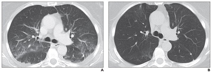
Fig. 11. 60-year-old man with metastatic pancreatic neuroendocrine tumor treated with everolimus.

A, Axial CT image (lung window) obtained 6 months after start of everolimus therapy when patient reported nonproductive cough shows newly apparent multifocal sub-pleural ground-glass opacities along bronchovascular bundles, likely representing mild drug-associated noninfectious pneumonitis.
B, Follow-up CT image obtained 3 months after everolimus cessation shows ground-glass opacities have resolved.
