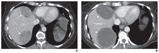
Fig. 6. 50-year-old woman with multiple liver metastatic lesions from pancreatic neuroendocrine tumor treated with sunitinib.

A, Axial contrast-enhanced CT image obtained before sunitinib treatment shows two solid masses (arrows) in liver.
B, Axial contrast-enhanced CT image obtained 6 months after sunitinib treatment shows masses (arrowheads) are larger. However, overall tumor attenuation and enhancing solid component have decreased, suggesting cystic change responding to treatment.
