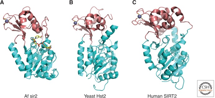
Figure 4.
Overall structure of sirtuins (Class III). Crystal structures of the sirtuin family of proteins are illustrated as cartoons highlighting the large Rossmann-fold domains (cyan) and small zinc-binding domains (brown). (A) Archaeoglobus fulgidus sir2 (PDB ID: 1ICI). NAD+ is drawn in stick model form in which yellow, blue, red, and orange represent C, N, O, and P atoms, respectively. (B) Yeast Hst2 (1Q14). (C) Human SIRT2 (1J8F). Zinc ions are represented as space-filled spheres in gray.

