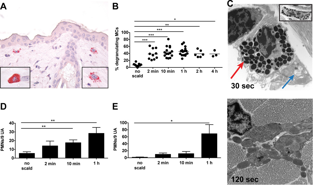
FIGURE 1.
MC degranulation is an immediate response to thermal challenge in WT mice. (A) Light micrograph of degranulating MCs in the skin as detected by CAE reactivity, 1 h after scald (magnification 630×). Left hand insert shows an intact MC, right hand insert shows a degranulating MC. (B) MC degranulation was quantitated in the skin at various times after the thermal challenge. The graph presents the percent of total MCs with >3 extracellular granules. Each dot is a separate mouse and the bar indicates mean value from 7 experiments. (C) EM of a degranulating MC captured 30 s and 120 s after thermal injury (original magnification 8,000×). At 30 s (top panel), there is segmental degranulation (blue arrow) and granules typical of a resting MC (red arrow). At 120 s (bottom panel), there is extensive MC degranulation (original magnification 12,000×). The top panel insert shows a fully granulated (intact) MC (original magnification 8,000×). (D, E) Quantitation of PMNs marginating (D) and extravasated (E) at various time points after thermal trauma. Data represent mean ± SEM, n=8–11 per time point from 7 experiments. *p < 0.05, **p < 0.01, ***p < 0.001.

