FIGURE 4.
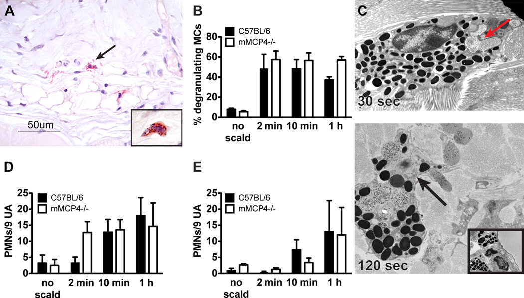
MC degranulation after thermal challenge is preserved in mMCP4−/− mice. (A) Histological demonstration (CAE reactivity) of degranulating MCs in the skin of an mMCP4−/− mouse after thermal challenge (arrow indicates extruded granules). Insert shows an intact MC from mMCP4−/− unscalded skin. (B) MC degranulation in mMCP4−/− mice is similar to that observed in WT mice. (C) EM of a mMCP4−/− MC captured 30 s after scald injury demonstrating zonal MC degranulation (arrow, top panel, original magnification 10,000×). By 120 s, prominent zonal MC degranulation is present (arrow, bottom panel, original magnification 10,000×). Insert shows an intact MC from mMCP4−/− unscalded skin (original magnification 8000×). (D, E) PMN margination (D) and tissue influx (E) in mMCP4−/− mice is comparable to WT controls. Data represent mean ± SEM, n=3–6 mice per time point from 3 experiments.

