FIGURE 8.
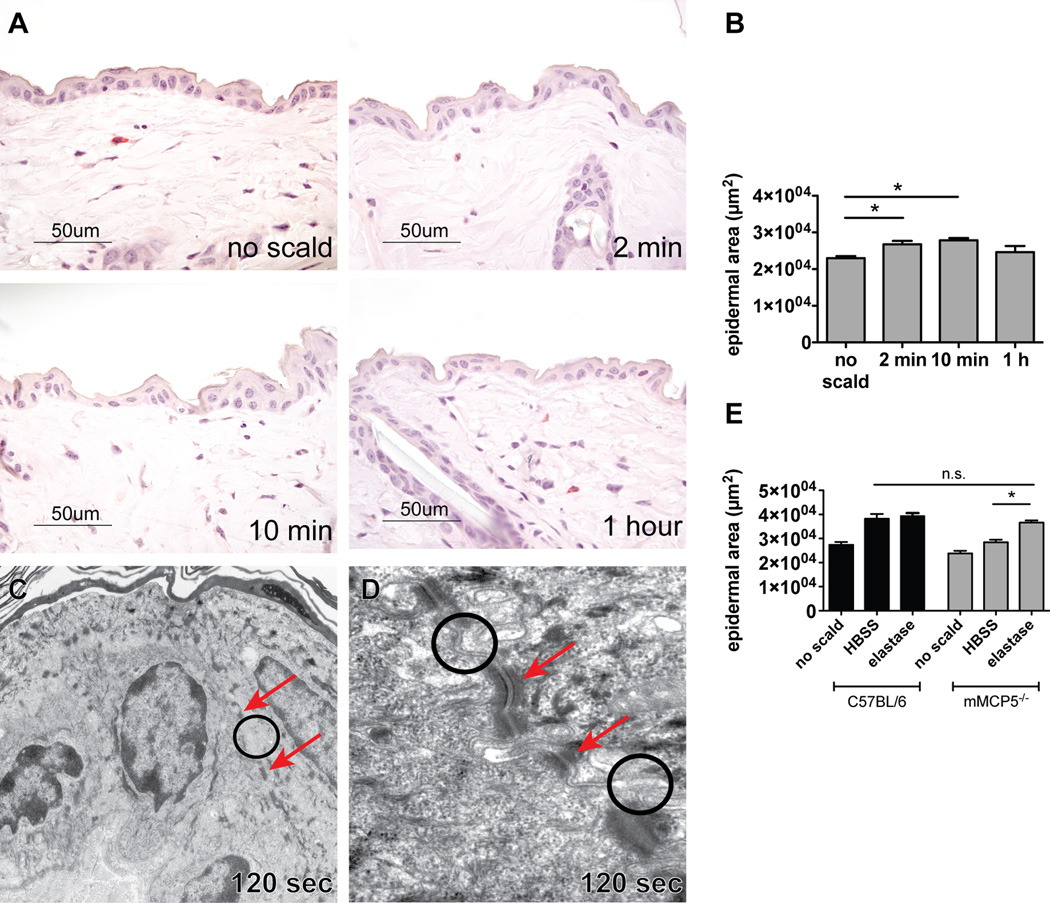
Thermal challenge of mMCP5−/− mice leads to mild epidermal changes. (A) Histology of normal mMCP5−/− mouse skin after shaving, no thermal injury and of thermally challenged mMCP5−/− skin 2 min, 10 min and 1 h after injury. (B) Epidermal area of unscalded and scalded skin measured 2 min, 10 min, and 1 h after thermal challenge of mMCP5−/− mice (mean ± SEM n=4–6 mice per time point from 3 experiments). (C) EM of mMCP5−/− mouse epidermis 120 s after thermal challenge showing normal appearance with intact TJs (circle) and desmosomes (arrows) (magnification 10,000×). (D) Higher magnification (60,000×) showing intact TJs (circles) and desmosomes (arrows) after thermal challenge. (E) Epidermal area in WT mice (black bars) and mMCP5−/− mice (grey bars) after application of HBSS or elastase to the scald area immediately after scalding, (mean ± SEM, n=3–5 mice per group from 2 separate experiments, mMCP5−/− no scald and WT elastase groups n=2)

