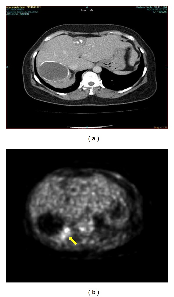
Figure 1.

The radiological findings of the patient. (a) Abdominal computed tomography showing a 65 × 55 mm stage 5 hydatic cyst in the right hepatic lobe and a 30 × 28 mm in the left hepatic lobe, (b) FDG18 positron emission tomography of the patient on which the mass revealed a high activity of FDG. Yellow arrow shows the high suvmax activity area (suvmax 80).
