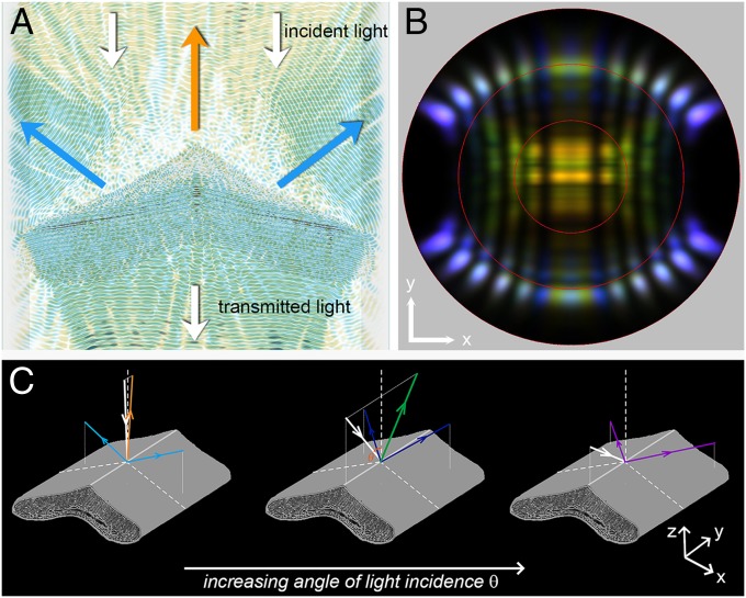
Fig. 4.
FDTD modeling of the breast feather barbule. (A) Two superimposed video frames for normal illumination of a breast feather barbule with 420 nm (blue) and 610 nm (orange) light. The time-lapse movies for these wavelengths are shown in Movie S2. The propagation direction of the incident and transmitted light is indicated by white arrows. The orange arrow indicates the propagation direction of the reflected orange light, and the blue arrow indicates the reflected blue light. (B) Calculated far-field light-scattering pattern for different incidence angles (0–60°), showing three-directional reflections for near-normal incidence; the intensity of the central reflection decreases for increasing angle of incidence (compare Fig. 3 B–D). The slightly different color of the two side beams reflected at the thin-film cortex is due to natural variations of the barbule thickness (Fig. S5). (C) Diagrams illustrating the angle-dependent reflection of the breast feather barbule for increasing angles of incidence θ in the longitudinal symmetry plane.

