Fig. 1.
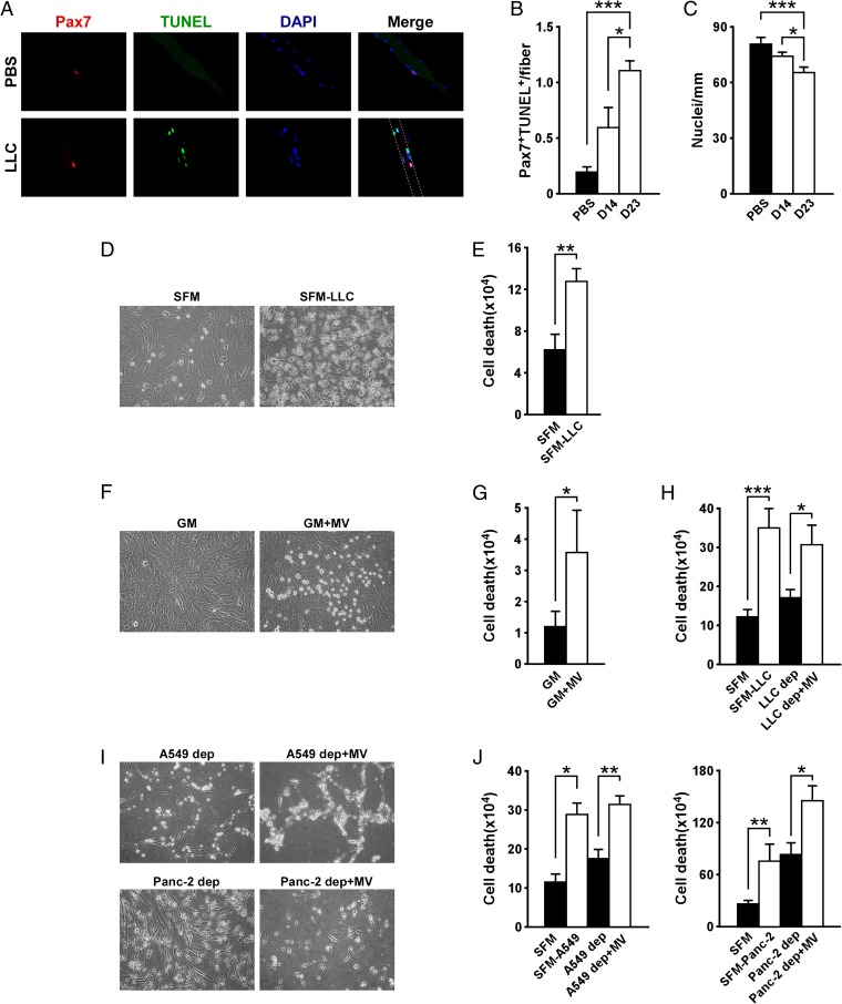
Tumor-derived MVs induce myoblast cell death in cancer cachexia. (A) Immunofluorescence performed on single myofibers isolated from in vivo xenograft LLC mouse models. As control, myofibers derived from PBS-injected mice were used. Pax7 is shown in red, TUNEL is shown in green, nuclei staining is in blue, and their colocalization is shown in the Merge panels. Staining was performed 14 and 23 d (D14 and D23, respectively) after tumor injection. (B) TUNEL quantitation related to immunofluorescence shown in A. (C) Determination of nuclei number per single myofiber. (D and E) Picture of C2C12 cells incubated with LLC-conditioned medium for 4 h (D). Serum-free medium (SFM) was used as a negative control. Cell death was determined with Trypan blue dye staining (E). (F–H) Picture of C2C12 cells incubated with LLC-derived MVs (F). Growth medium was used as negative control. Cell death was assessed with Trypan blue dye staining (G). The same assay was also performed on C2C12 cells incubated with MV-depleted medium (LLC dep) and LLC-derived MVs (LLC dep + MV) (H). (I and J) Induction of C2C12 cell death by A549- and Panc-2-derived MVs. Data are combined from at least three independent experiments. Results are presented as average ± SD. *P ≤ 0.05; **P ≤ 0.01; ***P ≤ 0.001.

