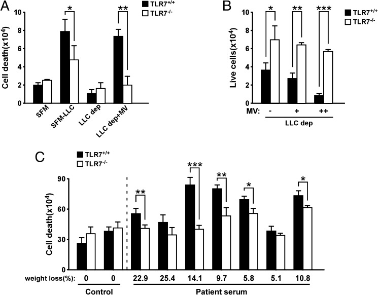
Fig. 3.
TLR7 is required for tumor-derived MV-induced cell death. (A) Trypan blue dye staining performed on primary myoblasts isolated from TLR7+/+ and TLR7−/− B6 mice and incubated with LLC-derived MVs for 48 h. As control, myoblasts incubated with serum-free medium (SFM), LLC-conditioned medium (SFM-LLC), and LLC MV-depleted medium (LLC dep) were used. (B) After 5 d of incubation with MVs, live cell number was determined by using a cell counter. MVs were resuspended in MV-depleted medium. “+” or “++” indicate a low or high amount of MVs being used to treat myoblasts. (C) Primary myoblasts isolated from TLR7+/+ and TLR7−/− B6 mice were incubated with MVs isolated from control serum of healthy donors (n = 2) or cachectic serum (n = 7) of pancreatic cancer patients with cachexia. Twenty hours after treatment, trypan blue dye staining was performed to assess for myoblast cell death. Results are presented as average ± SEM. *P ≤ 0.05; **P ≤ 0.01; ***P ≤ 0.001.

