
Fig. 3.
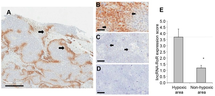
Linc-RoR is increased in hypoxic areas in vivo. HCC tumor cell xenografts were established in athymic mice to examine the expression of linc-RoR in vivo. Intratumoral hypoxic areas were identified by immunohistochemistry for Hypoxyprobe-1 (A). Scale bar: 500 µm. Immunohistochemistry for Hypoxyprobe-1 (B), in situ hybridization for linc-RoR (C) or in situ hybridization for negative control probe (D) in representative high-power fields from adjacent sections. Scale bars: 50 µm. The arrows show Hypoxyprobe-1- or linc-RoR-positive cells. (E) The number of linc-RoR-positive cells was quantified in hypoxic or nonhypoxic areas of tumor tissues. Data represents mean±s.e.m. of the number of positive cells with detectable linc-RoR in ten high-power fields. *P<0.05.