Fig. 8.
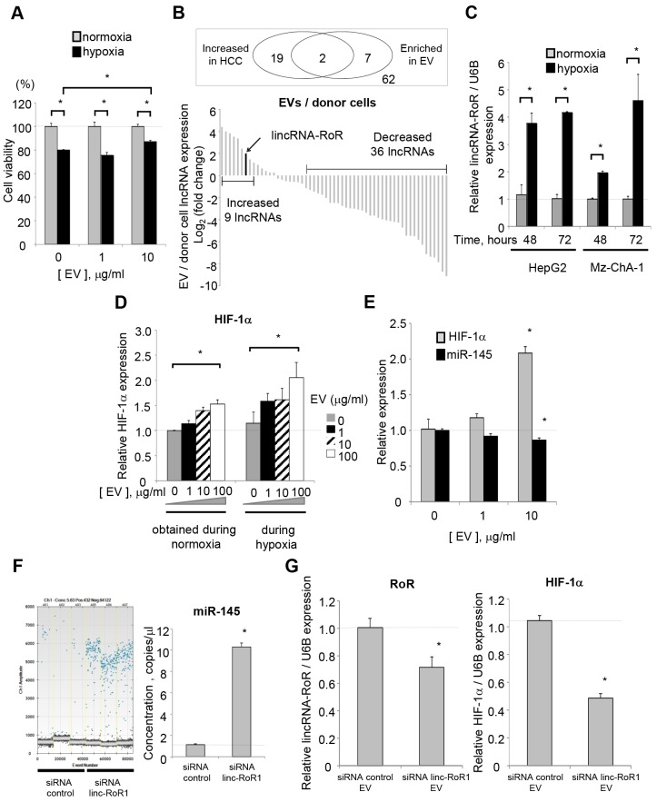
Extracellular linc-RoR during tumor cell responses to hypoxia. (A) HepG2 cells were transfected with siRNA 1 to linc-RoR. After 48 hours, cells were plated (1×10⁴/well) into 96-well plates in vesicle-depleted medium and incubated with varying concentrations of EVs under conditions of normoxia or hypoxia. Cell viability was assessed after 48 hours. (B) The expression of lncRNA, within EV preparations, released by HepG2 tumor cells from three independent replicates was assessed using LncProfilerTM qPCR Array Kit. Each bar represents the relative expression of extracellular RNA and donor-cell RNA for an individual lncRNA. Nine lncRNAs, including linc-RoR, were predominantly expressed in extracellular-RNA isolations compared to their donor cells. (C) Tumor cells were incubated under hypoxia or normoxia conditions, and extracellular RNA released by these cells was obtained after 48 or 72 hours. qRT-PCR for linc-RoR was then performed. (D) HepG2 cells were plated (1×10⁴/well) on 96-well amine-coated plates in vesicle-depleted medium and incubated with varying concentrations of EVs that had been derived from HepG2 cells under normoxia or hypoxia conditions. Recipient cells were then cultured under hypoxia conditions for 48 hours. Quantitative immunocytochemistry for HIF-1α was performed in recipient cells using an in-cell ELISA assay. (E) EVs were isolated from HepG2 cells under normoxia and added to recipient HepG2 cells. After 48 hours incubation under normoxia with those EVs, recipient-cell RNA was isolated and qRT-PCR for HIF-1α or miR-145 was performed. (F,G) HepG2 cells were transfected with siRNA 1 to linc-RoR, or nontargeting control, and cultured under normoxia. (F) After 72 hours, extracellular RNA from HepG2 cells was isolated and droplet digital PCR for miR-145 was performed. The number of positive droplets and concentration of miR-145 from three independent experiments are shown. (G) After 72 hours, EVs were collected from each group, and 10 µg/ml of EV was added to recipient HepG2 cells. After 48 hours incubation under normoxia, recipient-cell RNA was isolated and RT-PCR for linc-RoR or HIF-1α was performed.

