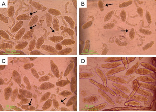
Fig. 3. : micrographic images of schistosomula cultured in 96 h: dead schistosomula showed an increased opacity and obvious lesions in the tegumental outer membrane. Sometimes the blebs appeared in their tegumental outer membrane (arrowed). A: schistosomula treated with 5 mg/mL Microtus fortis ( Mf )-albumin; B: schistosomula treated with 10 mg/mL Mf -albumin; C: schistosomula treated with represented 40% M. fortis serum; D: schistosomula treated with 10 mg/mL mouse serum albumin.