. 2013 Sep;108(6):741–754. doi: 10.1590/0074-0276108062013011

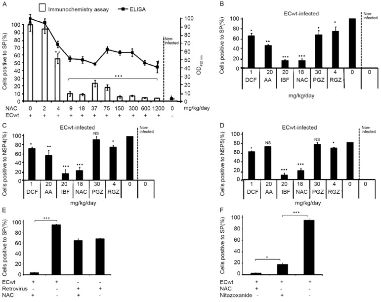
Fig. 2: effect of drug treatment on ECwt infection of villus intestinal epithelial cells (IEC). A: ICR mice were infected with ECwt and after 24 h post-inoculation (h.p.i.) mice were differentially subjected to treatment with increasing N-acetylcysteine (NAC) doses (n = 10 mice for each dose) during three days. Mouse small intestinal villi were isolated after three-day treatment and submitted to immunochemistry and ELISA analysis. Rabbit polyclonal antibodies (Abs) against rotavirus rotavirus structural proteins (SP), horseadish peroxidase (HRP)-conjugate goat anti-rabbit Ab and aminoethylcarbazole (AEC) substrate were used for immunochemistry analysis of methanol-acetic acid fixed villi. Guinea pig Abs against rotavirus SP for capturing rotavirus antigen and rabbit Abs against rotavirus SP for its detection were used for ELISA analysis of radio immunoprecipitation assay (RIPA) lysates from villus IEC. Reaction was revealed with HRP-conjugated goat anti-rabbit Ab and o-phenylenediamine dihydrochloride substrate and then read at optical density (OD) 492 nm . Villi or RIPA lysates of villi from both ECwt-infected and uninfected mice were used as control. Graph shows significant NAC inhibitory effect (p < 0.001); B: mice were infected with ECwt and treated from 24 h.p.i. with different drugs (n = 6 mice for each drug) at the indicated doses during three days. Immunochemistry analysis of villi was conducted as indicated in A for detecting rotavirus SP. Villi from ECwt-infected mice that had not been treated with drugs were used as a control; C: procedures were as indicated in B, except that rotavirus non-structural proteins NSP4 was detected in the immunochemistry ana-lysis; D: procedures were as indicated in B, except that rotavirus NSP5 was detected in the immunochemistry analysis. For results shown in A-D, villi isolated from either ECwt-infected or uninfected mice without treatment were used as a control. Results of drug treatment were found to be significant for B-D (p < 0.01); E: mice were infected with either ECwt or reovirus type 1 and after 48 h.p.i. mice (n = 4 mice for each experimental group) were treated with NAC (18 mg/kg/day) during three days. Rabbit Abs against reovirus SP, HRP-conjugated goat anti-rabbit Ab and AEC substrates were used for immunochemistry analysis of villi from reovirus infected mice. Graph shows significant NAC inhibitory effect (p = 0.0003); F: mice were infected with ECwt and after 24 h.p.i. mice (n = 4 for each experimental group) were treated with either NAC (18 mg/kg/day) or nitazoxanide (7.5 mg/kg/day) for three days. Graph shows significant NAC and nitazoxanide (NTZ) inhibitory effect in comparison to untreated infected control (p = 0.0003) and significant NAC inhibitory effect relative to NTZ treatment (p = 0.007). The immunochemistry analysis of villi was conducted as indicated in A for rotavirus SP. Data correspond to two independent experiments performed in duplicated and are expressed as percentage ± standard deviation (SD) of infected cells relative to total cells analysed in the case of the immunochemistry analysis or mean percentage ± SD of the rotavirus antigen present in RIPA lysates of villi from infected and NAC-treated relative to that from infected and untreated mice. AA: ascorbic acid; DCF: diclofenac; IBF: ibuprofen; PGZ: pioglitazone; RGZ: rosiglitazone.

Fig. 2: