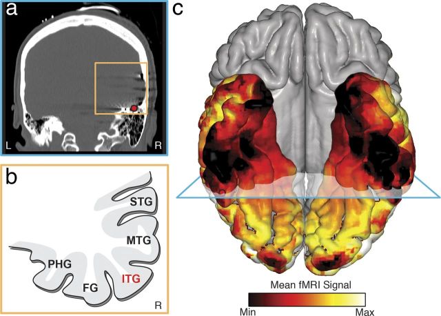
Figure 5.
fMRI eclipse zone. a, CT image of a subject's brain in the coronal plane showing a strip of electrodes under the temporal lobe. The electrode with preferential response to numerals is marked with a red circle. There is air-filled petrous bone located underneath the electrode. b, Schematic of the gyri surrounding the ITG. MTG, Medial temporal gyrus; STG, superior temporal gyrus. c, The mean fMRI signal in 6 healthy control subjects was normalized to a range of 0–1 for each individual and then averaged across subjects and rendered on a standard MNI brain. The area with a preferential response to numerals indicated by a red circle falls nearby the core of the signal dropout. c, Blue plane represents the coronal plane for a and b.

