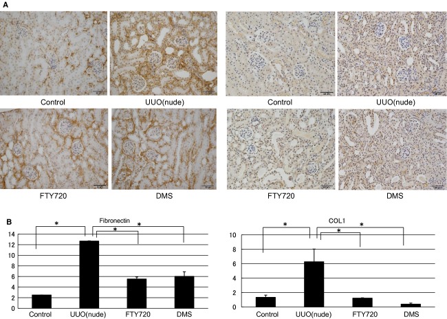
Figure 11.
Examination of immunohistochemical staining in the UUO of nude mouse model. Immunohistochemistry showed that the stained area was expanded in the left kidney compared with the control right kidney in the nude mice. In the kidney of the nude UUO mouse model, FTY720 and DMS pretreatment significantly decreased the staining intensity of fibronectin and COL1. N = 3. Data are presented as means ± SE. *P < 0.05. UUO, unilateral ureteral obstruction; DMS, N,N‐dimethylsphingosine; COL1, collagen type 1.

