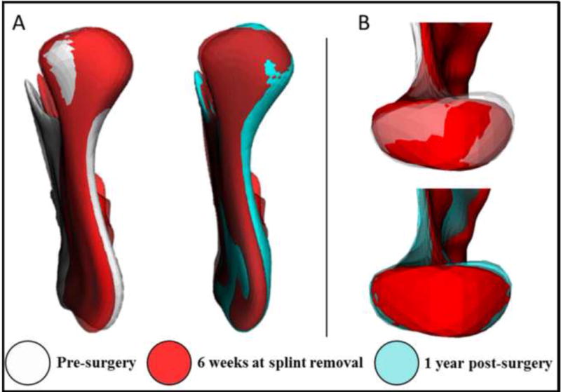
Fig. 8.

Two-jaw surgery patient. A: posterior view of the ramus showing lateral displacement. B: superior view of the condyle of the same patient showing its rotation. Note that the medial pole is displaced more than the lateral pole for surgical movements and post-surgical adaptation.
