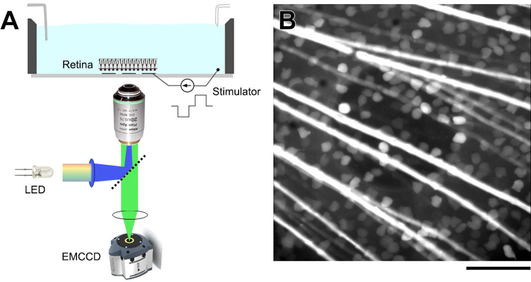
Figure 1.
(A) Experimental setup used to image RGC calcium fluorescence in ex vivo salamander retina. Images were captured with an electron-multiplying CCD camera during stimulation with a single MEA electrode. (B) Fluorescence image of a salamander retina loaded with OGB-1 dextran and mounted on a transparent MEA. RGC somata and axon bundles are clearly visible. A 200-µm-diameter indium-tin-oxide electrode is centered in the field of view. Scale bar is 100 µm.

