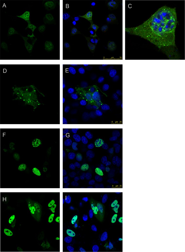
Figure 4. Foci forming analysis of the CT2AXH region.
Residues TLND to AXH from ataxin-1 in fusion with YFP forms small foci that are extranuclear (A and B). One cell from the image is enlarged in C for clarity. N terminal extension of this region where the construct started after the polyQ tract (CT2AXH) formed larger foci which also were extranuclear (D and E). Addition of an NLS to this construct resulted in nuclear foci formation (F and G). Deletion of CT2AXH from ataxin-1 resulted in diffused expression of the protein (H and I). A, D, F and H show YFP fluorescence. YFP fluorescence is overlaid with DAPI in B, C, E, G and I.

