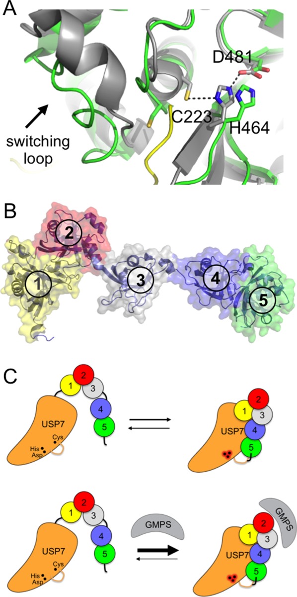
Figure 2.

Activation of USP7 by the HUBL domain. A: Active site of USP7 in the presence (gray) and absence (green) of bound ubiquitin aldehyde (yellow; 1NB8 and 1NBF). Arrow indicates switching loop. B: HUBL domain of USP7, consisting of five UBL domains (numbered). C: Model for activation of USP7 by the HUBL domain, which is promoted by GMPS binding. The HUBL 4 and 5 domains bind to USP7 and trigger a conformational change in the switching loop, which in turn results in a reconfiguration of the catalytic triad to the active conformation. GMPS binding stabilizes the association of the HUBL-4, 5 domains and thereby maintains the active state. Adapted from Ref.36.
