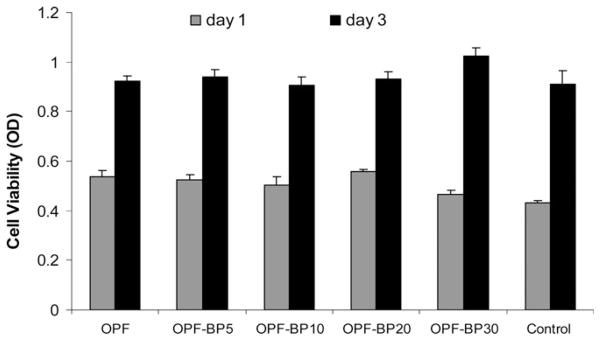
Fig. 5.

Toxicity of leaching materials from hydrogels after 1 and 3 d in culture. hFOB cells were plated on 24-well tissue culture plates and exposed to leaching materials from scaffolds via transwell inserts. The viability of the cells was tested using the MTS Cell Proliferation Assay by measuring the optical density (OD) at 490 nm. Values represent mean ± standard error (n = 3).
