
Fig. 10.
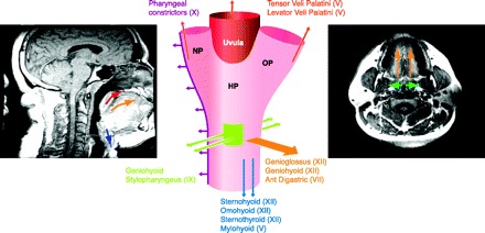
The upper airway in obstructive sleep apnea: a reliance on upper airway dilator muscles for patency. Magnetic resonance images sagittal (left) and coronal (right) of a subject with obstructive sleep apnea. The airway is narrowed but remains patent in wakefulness, in large part because of key dilator muscles, labeled in the center diagram with cranial nerve innervations in parentheses. Arrows indicate overall force vector and are shown on both the diagram and images. Upward directed arrows (red) signify force vectors for levator palatini and tensor veli palatini muscles in raising the soft palate (uvula) and lateral walls. Because the pharynx is collapsible at all tangents, multiple muscle groups must act in concert to prevent collapse of the pharynx.