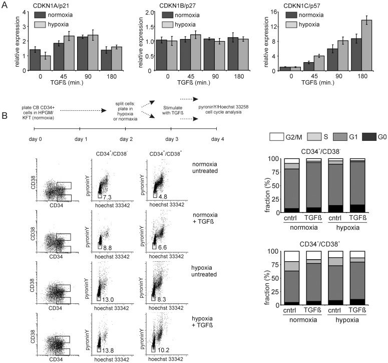
Figure 3. p57 and HSC quiescence are induced by both TGFβ and hypoxia.
A. CB CD34+ cells were cultured under normoxic and hypoxic conditions for 24 hours and stimulated for different times with 1 ng/ml TGFβ where after Q-RT-PCR was performed for CDKN1A/p21, CDKN1B/p27 and CDKN1C/p57. A representative experiment out of 2 independent experiments is shown, error bars indicate standard deviation of PCR performed in triplicate. B. CB CD34+ cells were treated as indicated in the scheme and cell cycle analysis was performed using Hoechst and PyroninY staining. Representative FACS plots and the cell cycle distribution in CD34+/CD38− and CD34+/CD38+ fractions are shown.

