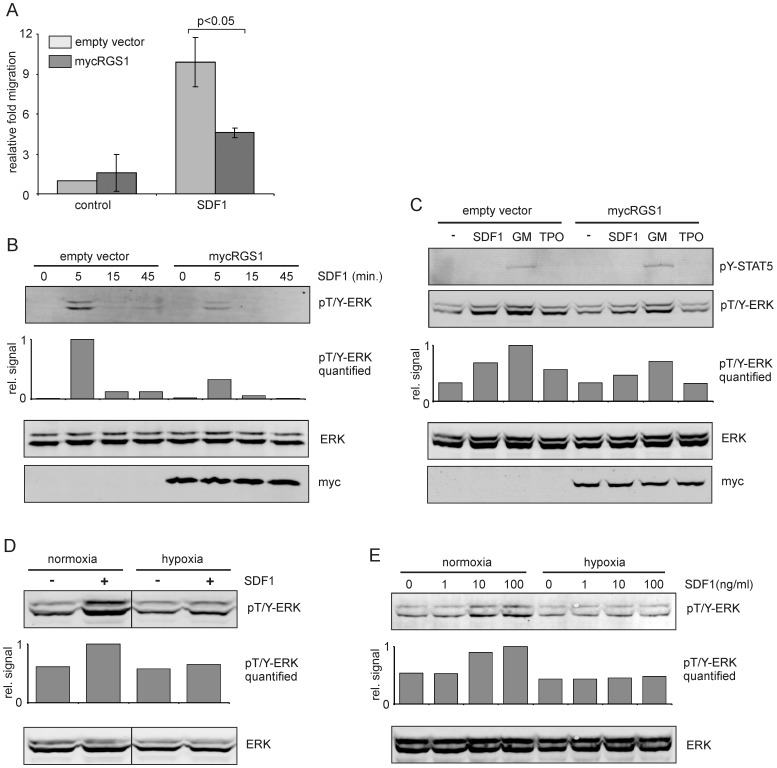
Figure 6. RGS1 dampens SDF1-induced migration and signaling.
A. Transwell migration assay towards SDF1 (100 ng/ml) using CB CD34+ cells transduced with empty vectors or RGS1. The mean and standard deviation of three independent experiments is shown. B. Western blot of CD34+ cells transduced with empty vectors or RGS1 that were stimulated with SDF1 (20 ng/ml) for different timepoints. C. Western blot of OCI-AML3 cells transduced with empty vectors or RGS1 that were stimulated with different cytokines (20 ng/ml each) for 10 minutes. D. OCI-AML3 cells were cultured for 24 hours under normoxia or hypoxia, stimulated with 20 ng/ml SDF1 for 10 minutes and Western blotted for phosphorylated ERK1/2. E. UT7-GM cells were cultured for 24 hours under normoxia or hypoxia, stimulated with 20 ng/ml SDF1 for different timepoints and Western blotted for phosphorylated ERK1/2. Band intensities for phospho-ERK1/2 were measured and plotted as relative signal (in B-E). Representative experiments out of 2-3 independent experiments are shown.

