
Figure 4.
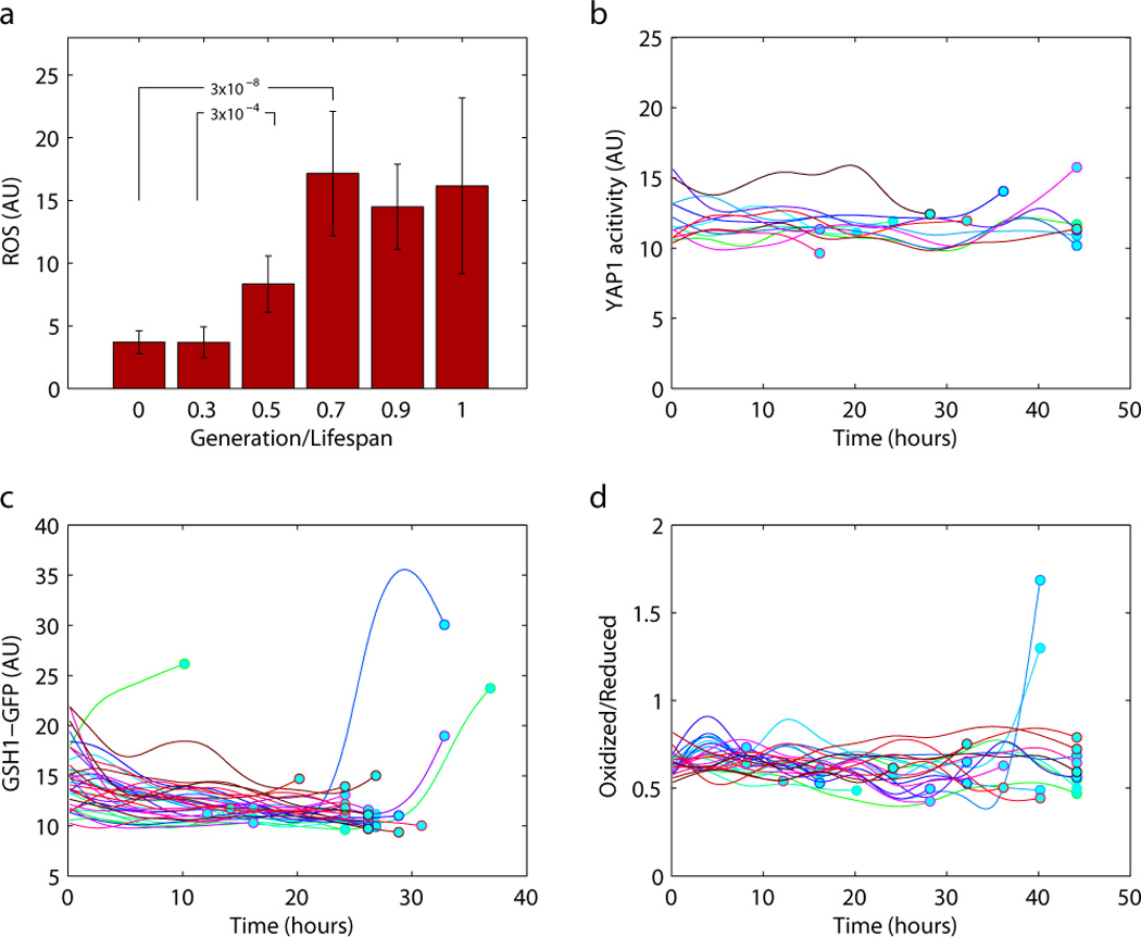
Oxidative stress as a function of age in individual cells. a) DCFH-DA staining showed increased ROS level in the aged cell populations. Staining was performed at 0, 11, 22, and 33 hours after cell loading in separate channels. Cells were grouped by their relative lifespan (generation normalized by the total lifespan) at the time of staining. b) Yap1 activity as a function of age remains the same throughout the lifespan. Yap1 activity is reported by a YFP driven by a crippled CYC1 promoter containing a Yap1 binding site. c) The protein level of Gsh1 (a transcriptional target of Yap1) as reported by a 3’ GFP fusion does not increase with age. d) Redox potential reported by the roGFP. The ratio of the oxidized to the reduced forms of the roGFP (arbitrary unit) was calculated from the fluorescence signals from the two different forms, as excited by two different wavelengths.