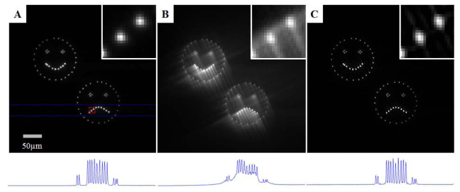
Fig. 4.

Panels A-C: Comparison of the focal plane images from (panel A) a conventional microscope, (panel B) the raw extended DOF image and (panel C) the restored extended DOF image. Note the contrast of these three images has been enhanced to have 0.1% of the pixels saturated to aid in visualization. Outlined in red in panel A is a region containing two points from the frown in the bottom right feature. A close up of this feature is given in the upper right region of each panel to provide a detailed examination of the imaging quality. Outlined in blue in panel A is a region containing 14 points. The maximum intensity projection of this region is shown below each panel showing that the digital restoration yields contrast similar to the conventional image.
