
Fig. 6.
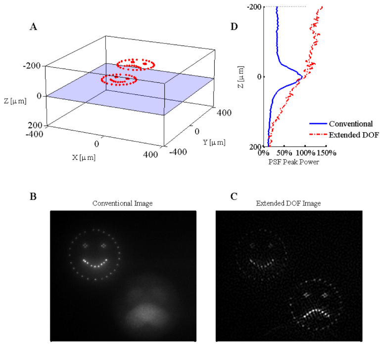
Experimental results for the three-dimensional SLM illumination in scattering media with both the conventional and extended depth of field microscope. The three-dimensional illumination pattern is shown in panel A. The focal plane is located 220µm below the surface of the scattering media. The relative intensity of the fluorescence as a function of depth is given to the right of panel A. The results from imaging the three-dimensional pattern in bulk fluorescent material are given for the conventional microscope (panel B) and the extended DOF microscope (panel C). Contrast was enhanced to saturate 0.02% of the pixel values in panels B and C ( Media 3 (6.6MB, AVI) ).