
Figure 5. Expression of cell proliferation markers was increased in the hearts of R2M mice during postnatal growth.
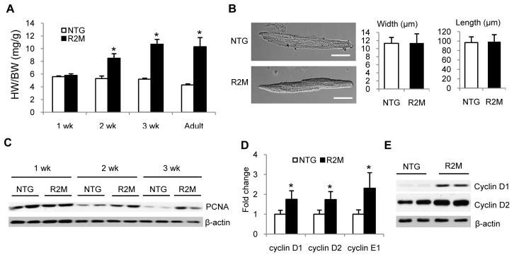
(A) HW/BW of R2M mice at 1, 2, 3 weeks (wk), and 2 months (adult). (B) Left, typical cardiomyocytes from NTG or R2M mouse hearts at 2 weeks. Right, average length and width of cardiomyocytes isolated from 2 week old NTG and R2M hearts. A total of 100–120 cardiomyocytes were counted in each heart, n=3 for each genotype. Scale bar, 20 μm.(C) Representative immunoblot of PCNA in whole heart lysates of NTG and R2M mice at 1, 2 and 3 weeks. (D) mRNA and (E) protein levels of cyclin D1, D2 and E in whole heart lysates of NTG and R2M mice at 2 weeks. n=4; *p< 0.05 versus NTG.