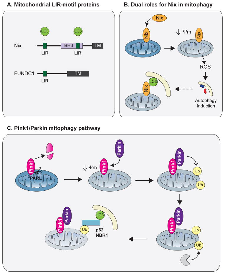
Figure 5. Mitophagy.
(A) Domain architecture of the mitophagy adaptors Nix and FUNDC1. Both proteins contain a transmembrane domain embedded in the outer mitochondrial membrane (OMM) as well as a LIR, which mediates recruitment of APs to mitochondria. (B). Following mitochondrial stress, Nix promotes mitochondrial depolarization and ROS formation, leading to autophagy induction. Nix also mediates targeting of APs to mitochondria via its LIR. (C) In healthy polarized mitochondria, PINK1 is constitutively degraded by the inner mitochondrial membrane protease PARL. Following depolarization, full length PINK1 accumulates on the OMM and recruits the E3 ligase Parkin. Parkin ubiquitinates multiple OMM proteins, leading to proteasomal degradation of OMM components and p62/NBR1-mediated AP recruitment to mitochondria. Abbreviations: TM, transmembrane domain; Ψm, mitochondrial membrane potential; ROS, reactive oxygen species.

