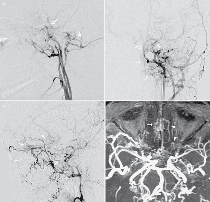
Figure 3.
Left carotid artery angiogram (A,C: LCCA in lateral and AP view; B: LECA in lateral view) and MIP images of TOF-MRA (D). The left ICA was also hypoplastic beginning from the carotid bifurcation, the 5th and 6th segments were tapered, and the 7th segment was mainly supplied by the reverse flow from left PComA (thick arrow). Abundant thin arterial collaterals from sphenopalatine, inferior orbital and angular branches of the left IMA were observed in the nasal cavity, ethmoidal sinus and ethmoidal planum (thin arrow). The A2 segment of the left ACA was also opacified by those collaterals through their anastomoses with a fronto-orbital branch of the left ACA. Note that the fronto-orbital artery made a peculiar “hair-pin” turn (arrowhead) in its proximal segment. A hypoglossal branch of the left ECA also supplied the BA (open arrow) with another intradural network formation (asterisk).

