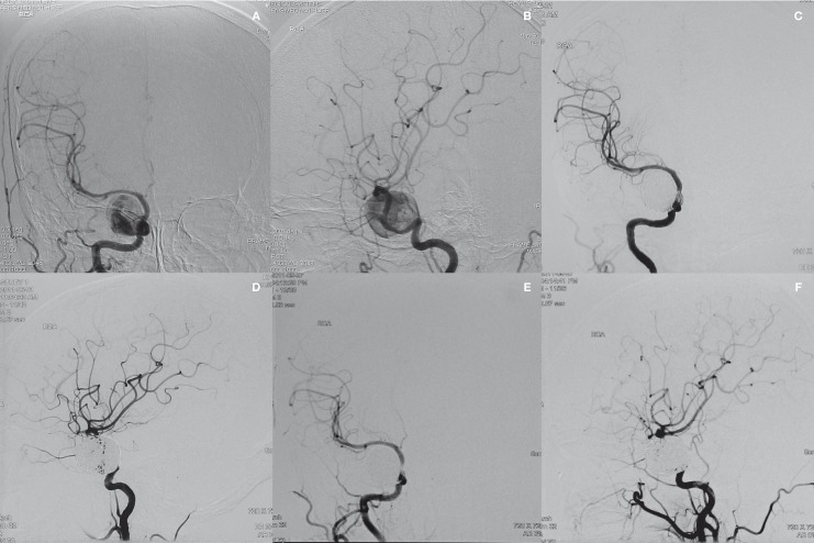
Figure 1.
Right carotid angiogram on anteroposterior (A) and lateral (B) views from a 57-year-old woman with diplopia demonstrating a giant intracavernous carotid artery aneurysm. After 2 sessions of stent/coil embolization, right carotid angiogram on anteroposterior (C) and lateral (D) views demonstrating complete obliteration of the aneurysm. Right carotid angiogram on anteroposterior (E) and lateral (F) views at 3 months postembolization showing complete obliteration of the aneurysm.

