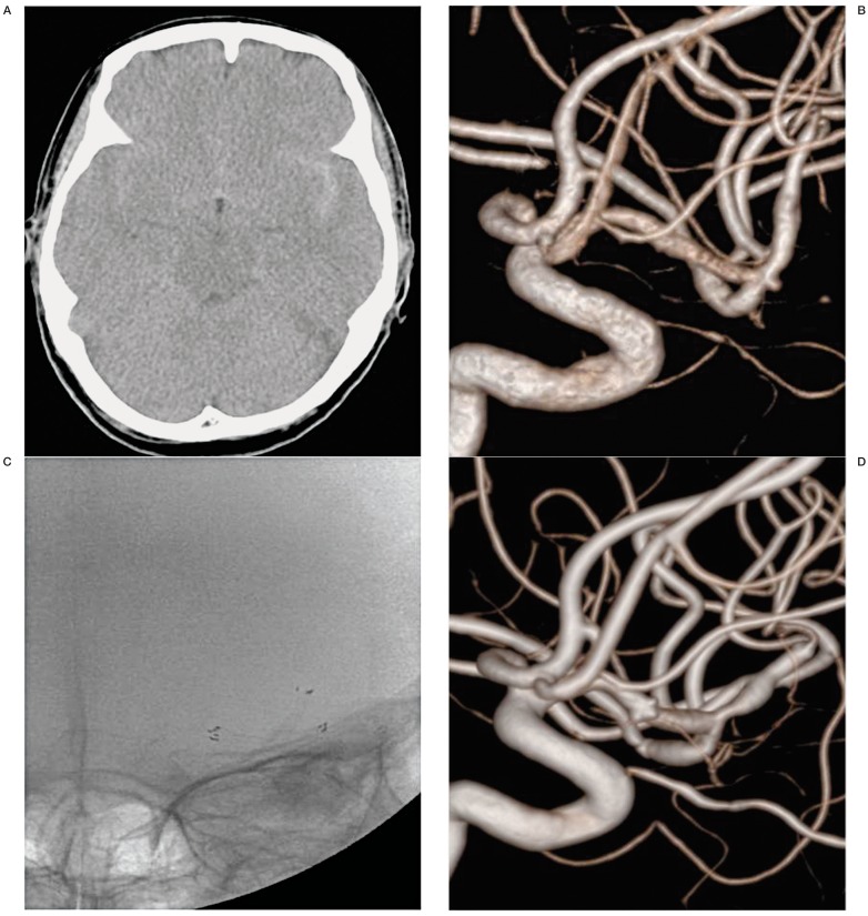
Figure 1.
A 70-year-old woman (Case 1) with a ruptured MCA aneurysm. A) Subarachnoid hemorrhage in basal cistern and predominantly left sylvian cistern on the brain CT scan. B) Very small aneurysm (1.3 × 1.1mm) at the MCA bifurcation on 3-dimensional rotational angiogram. C) Y-configured stenting for aneurysm of MCA bifurcation on the fluoroscopy. D) A follow-up angiogram 22 months later demonstrated the disappearance of the aneurysm and the presence of asymptomatic post-stent stenosis at both M2 segments.

