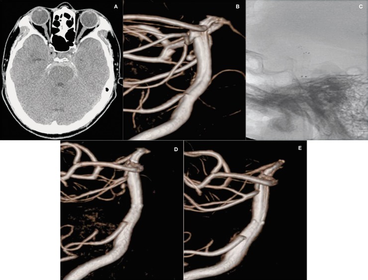
Figure 2.
A 52-year-old man (Case 5) with a ruptured aneurysm on the dorsal wall of the basilar trunk. A) Subarachnoid hemorrhage in the prepontine cistern on the brain CT scan. B) Very small aneurysm (2.1 × 2.0 mm) of the basilar trunk on the dorsal wall on 3-dimensional rotational angiogram. C) Double stents using the stent-within-stent technique on fluoroscopy. D) Decreased contrast filling within the aneurysm on the immediate post-operation angiography. E) Follow-up ICA angiogram at 18 months post-operation shows the disappearance of the aneurysm.

