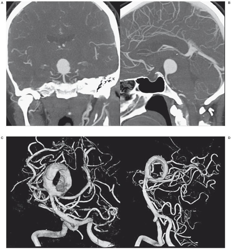
Figure 1.
Incidental large basilar tip aneurysm in a 50-year-old woman. A,B) CT angiography demonstrates large spherical basilar tip aneurysm. C,D) 3DRA of the same aneurysm 10 days after stent placement in the right posterior cerebral artery. The aneurysm is now largely thrombosed with a donut-shaped remaining lumen.

