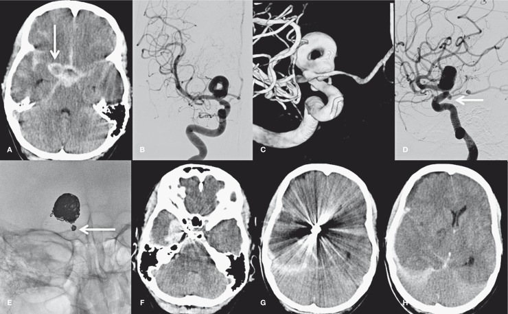
Figure 3.
A 46-year-old man with poor grade subarachnoid hemorrhage. A) CT scan shows SAH and aneurysm (arrow). B-D) 2D angiography (B,D) and 3DRA (C) demonstrate a donut-shaped large carotid tip aneurysm and a second aneurysm on the PcomA (arrow in D). E) Coil meshes after coiling of the carotid tip aneurysm and the small PcomA aneurysm (arrow). F-H) CT scan 10 days after coiling shows recurrent hemorrhage from the PcomA aneurysm with a large subdural component and mass effect.

