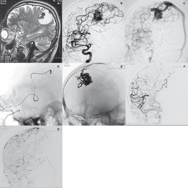
Figure 1.
Patient 2. A) MRI scan demonstrated abnormal vascular structures at the right central area. B,C) Angiogram showed a fronto-parietal AVM, supplied by two branches of the fronto-parietal ascending arteries and drained by a superficial vein into the superior sagittal sinus. D) Onyx embolization by CAIT was performed. E) Plain radiography showed the final cast of Onyx. F,G) Post-procedural angiogram demonstrated complete exclusion of the AVM.

