
Figure 2.
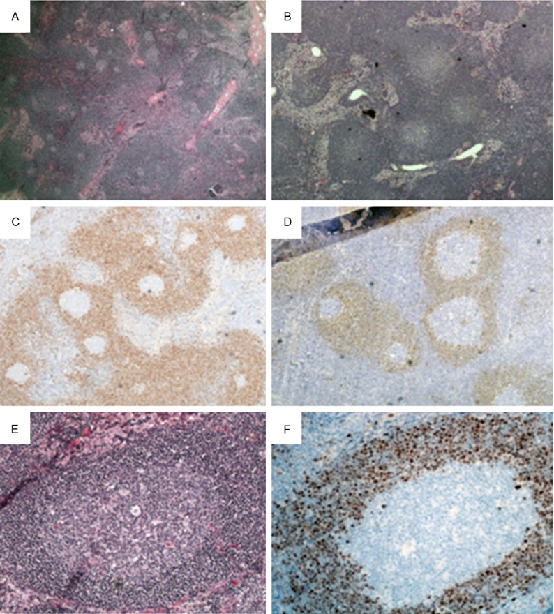
MCL-MZGP in lymph nodes. H&E staining in case 5 (A, X20) and case 4 (B, X40) reveals numerous reactive-appearing follicles with germinal centers and focally intact sinuses. Immunohistochemical staining for cyclin D1 highlight expanded mantle zones in case 5 (C) and case 4 (D) (X40). (E) Higher power view of one reactive-appearing follicle with germinal centers and expanded mantle zones (H&E, X200). (F) Higher power view of cyclin D1 staining in one follicle (X200).