
Figure 3.
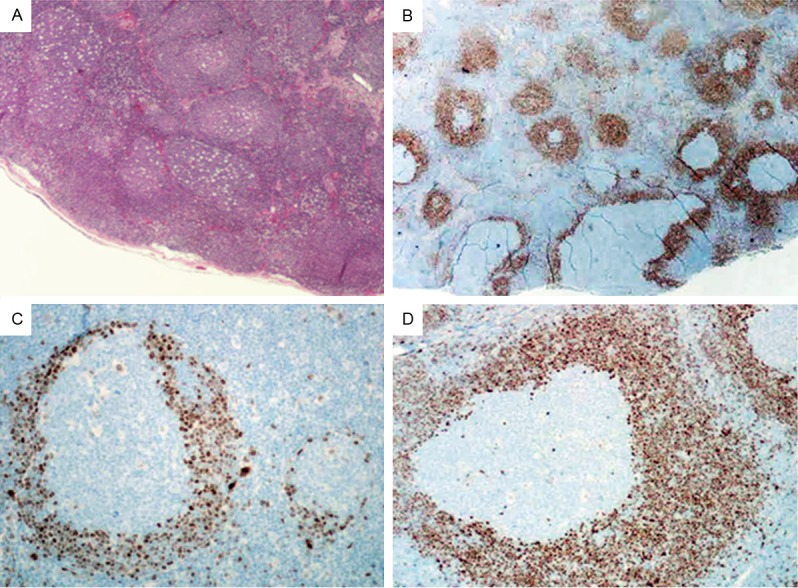
Histopathology and immunohistochemical studies in case 3. (A) H&E staining reveals numerous reactive-appearing follicles (X40). (B) immunohistochemical staining for cyclin D1 reveals a mixture of MCL-MZGP and “in situ MCL” (X40). Higher power view of Cyclin D1 staining for “in situ MCL” (C) and MCL-MZGP (D) (X200).