
Figure 2.
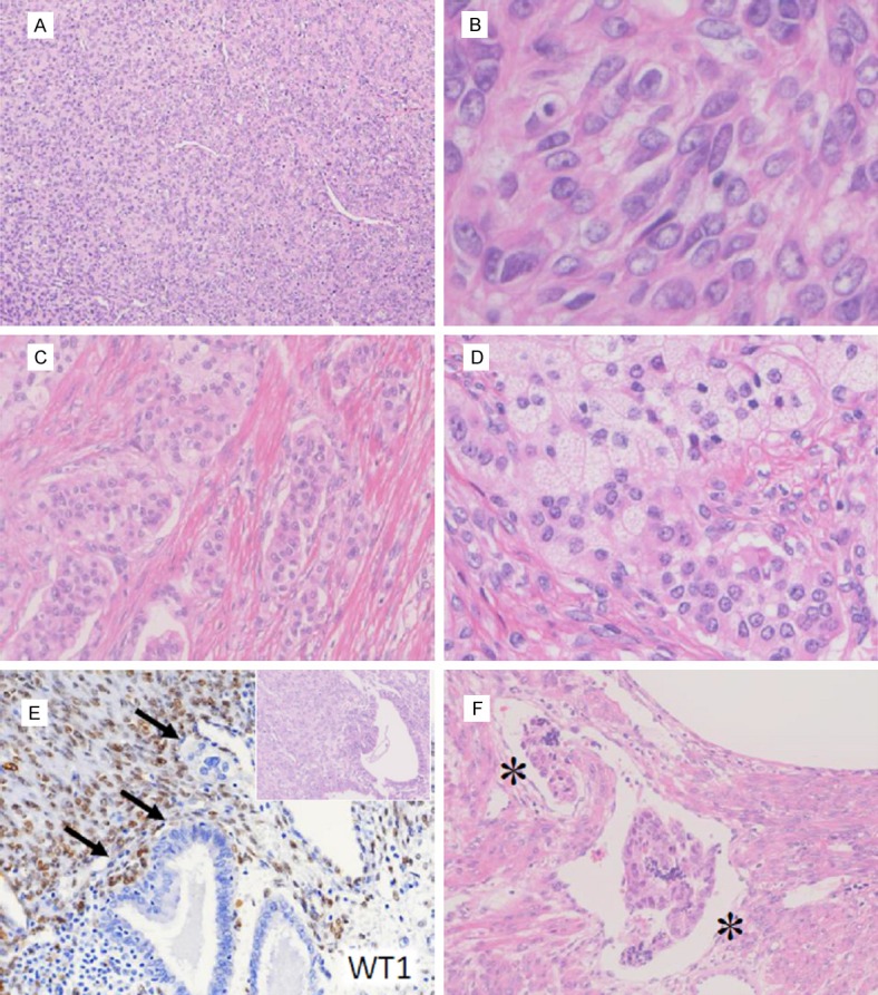
Microscopic features of Case 2. (A, B) The solid area composed of epithelioid cells (A). High-power field of diffusely proliferating tumor cells are shown (B). (C, D) Epitheliod cell nests with sex cord-like differentiation (C) with lipidized cells (D) are shown. (E) Endometrial glands are infiltrated by WT1-positive tumor cells (arrows). Inset: HE staining of the serial section. (F) Vascular space invasion is detected in the tumor periphery. The asterisks indicate the vessel lumen.