
Figure 1.
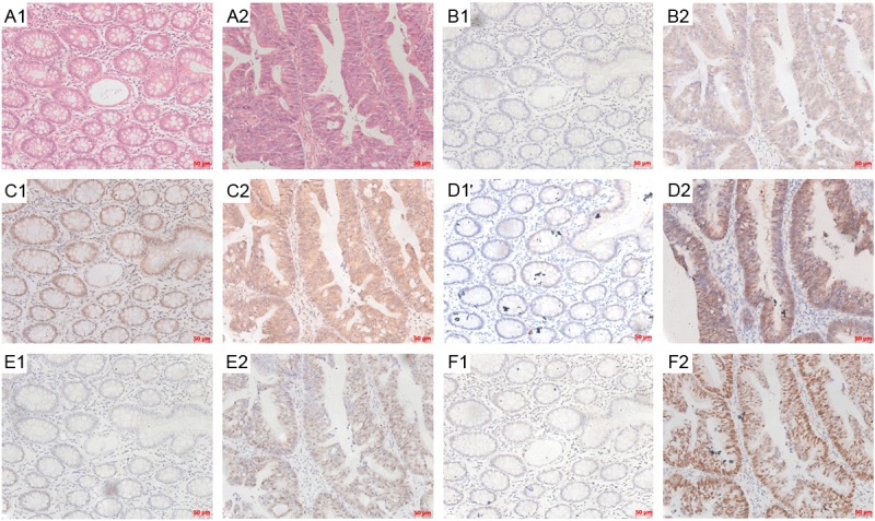
Immunohistochemical staining results for Nrf2, NQO1, MRP1, cmyc and p53 expression in CRC. The unstained tissue slides from CRC were subject to HE staining (A1, A2) and IHC staining with antibodies against cmyc (B1, B2), MRP1 (C1, C2), NQO1 (D1, D2), Nrf2 (E1, E2) and p53 (F1, F2). Adjacent normal colorectum tissues were shown in A1, B1, C1, D1, E1 and F1 panels, while CRC tissues were shown in A2, B2, C2, D2, E2 and F2 panels. The bar scales were labeled at the lower right corner of each pictures.