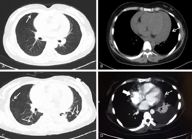
Figure 1.
Images of the chest computed tomography (CT) on admission (A and B) and enhanced CT in a few days (C and D). (A and B) A scattered nodule in the lungs (arrow), and severe pericardial effusion (dashed arrow); (C and D) New scattered nodules in the lungs (arrows), mild pericardial effusion (dashed arrow), and a mass suspected in the right atrium (block arrow).

